









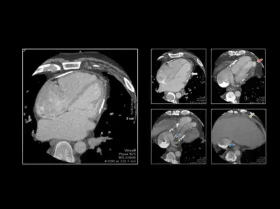










A prospective study of 79,163 post-contrast CT examinations has found that 40.7% of superior mesenteric artery occlusions (SMAO) were missed on the initial interpretation. The research, published in Clinical Imaging and conducted by vRad, identified 83 missed cases over six weeks across 766 imaging facilities. Each was confirmed through quality assurance review and the report addended within one hour of the original read.
Key findings
- 40.7% of SMAO cases were missed on the initial interpretation.
- SMAO is rare, with a 0.26% incidence in the study population.
- None of the missed cases had SMAO or mesenteric ischemia noted in the clinical indication, compared with 17.4% of cases diagnosed correctly on the first read.
- Only 2.4% of QA-detected cases were CT angiography exams, versus 46.3% of cases caught on the initial read.
- The AI model demonstrated 67.6% sensitivity and 99.6% specificity in prospective deployment.
- Model-positive exams reached a radiologist in a median of 5.1 minutes, compared with 27.9 minutes for model-negative exams.
How the AI quality assurance model works
A quality assurance AI model analyzed each exam in the study and flagged cases in which SMAO was suspected but not mentioned in the original radiology report. Those flagged examinations were then reviewed by a second radiologist as part of the quality assurance process. Through this approach, researchers confirmed 83 cases of SMAO that had been missed on the initial interpretation, representing 40.7% of all cases identified during the study period.
The SMAO model is one of 19 pathologies that run automatically on all relevant examinations entering the vRad system, providing worklist prioritization and an AI QA safety net for vRad reading radiologists. External radiology practices using The vRad Platform also benefit from these AI models in the form of worklist prioritization.
AI does not replace the radiologist’s search pattern or diagnostic judgment. For a rare and easily overlooked finding, it provides an additional opportunity to identify a consequential result when it is missed on the initial search.
How the model was evaluated
- Deployed prospectively across 79,163 post-contrast CT exams with abdominal coverage.
- Drawn from 766 imaging facilities over a six-week study period.
- Flagged exams where SMAO was suspected but not mentioned in the original report.
- 237 exams were flagged for review; 83 were confirmed as missed SMAO.
- Each confirmed case was reviewed and the report addended within one hour of the initial read.
Why is SMAO missed on CT?
SMAO is rare — the study measured a 0.26% incidence — and easy to overlook. When the vessel is fully occluded, its attenuation can approximate that of the surrounding mesentery, leaving little visual cue to draw the eye. In early acute cases, the rest of the abdomen and pelvis may appear unremarkable apart from a small focal thrombus.
The study points to search pattern rather than recognition failure. Not one of the QA-detected cases had SMAO or mesenteric ischemia mentioned in the clinical indication, compared with 17.4% of the cases diagnosed correctly on the first read. QA-detected cases were also far less likely to be CT angiography exams: 2.4%, against 46.3% of initially diagnosed cases.
Taken together, these findings suggest the superior mesenteric artery is evaluated less consistently when nothing in the order prompts it, and that the more subtle intensity changes on conventional post-contrast CT make the diagnosis harder when the vessel is assessed at all. The implication is that this is a correctable gap in how the exam is searched, not a limit on what radiologists are able to recognize.
Key takeaways from the study team
The study was led by Robert J. Harris, Director of Imaging AI at vRad. Its authors conclude that search pattern, rather than an inability to recognize SMAO once the vessel is carefully assessed, is the principal contributor to missed cases.
The research also evaluated the AI model itself. Deployed prospectively, it demonstrated 67.6% sensitivity and 99.6% specificity. Exams with a positive model result reached a radiologist substantially faster, with a median delay of 5.1 minutes compared with 27.9 minutes for model-negative exams.
Read the full study in Clinical Imaging (2026;134:110810).
Key takeaways from vRad Chief Medical Officer, Benjamin W. Strong, MD
Radiology practices may benefit from specifically encouraging radiologists to include the SMA more consistently in their search pattern, said vRad Chief Medical Officer Dr. Benjamin Strong, particularly on conventional post-contrast CT where the intensity changes are more subtle than on CTA.
Summary
A prospective study of 79,163 post-contrast CT examinations, published in Clinical Imaging and conducted by vRad, found that 40.7% of superior mesenteric artery occlusions were missed on the initial read. The study attributes the misses primarily to search omission rather than difficulty recognizing the finding, based on the near-total absence of SMAO in the clinical indications of missed cases and their low rate of CT angiography.
For radiology practices, the actionable conclusion is that more consistent evaluation of the superior mesenteric artery on conventional post-contrast CT may reduce missed cases. AI adds a second layer: the SMAO model is one of 19 pathologies running automatically on all relevant examinations entering the vRad system, providing worklist prioritization and an AI QA safety net for vRad reading radiologists, with external practices using The vRad Platform benefiting from these models in the form of worklist prioritization.
Frequently asked questions
How often is superior mesenteric artery occlusion missed on CT?
In a prospective study of 79,163 post-contrast CT examinations published in Clinical Imaging, 40.7% of superior mesenteric artery occlusions were missed on the initial interpretation and identified later through AI-assisted quality assurance review.
Why is SMAO difficult to detect on CT?
When the superior mesenteric artery is fully occluded, its attenuation can approximate that of the surrounding mesentery, leaving little visual cue. In early acute cases the rest of the abdomen may appear normal apart from a small focal thrombus. The study also found that missed cases were rarely CT angiography exams and almost never carried a clinical indication pointing at SMAO, suggesting search omission is the larger factor.
How common is superior mesenteric artery occlusion?
The study measured an incidence of 0.26% across 79,163 post-contrast CT examinations with abdominal coverage.
Can AI detect superior mesenteric artery occlusion?
In this study, a deep learning model deployed prospectively demonstrated 67.6% sensitivity and 99.6% specificity for SMAO. Used as a quality assurance safety net, it identified 83 cases that had been missed on the initial read over a six-week period.
Is the SMAO AI model available to other radiology practices?
The SMAO model is one of 19 pathologies that run automatically on all relevant examinations entering the vRad system. External radiology practices using The vRad Platform benefit from these AI models in the form of worklist prioritization.
Where was this study published?
The study was published in Clinical Imaging (2026; volume 134, article 110810) and led by Robert J. Harris, Director of Imaging AI at vRad. The full text is available at doi.org/10.1016/j.clinimag.2026.110810.