Quality improvement initiative to increase radiologist reporting of the Alberta stroke program early CT score (ASPECTS)
Journal Name: Clinical Imaging
Published: 11/01/2025
Remote radiologist jobs with flexible schedules, equitable pay, and the most advanced reading platform. Discover teleradiology at vRad.

Radiologist well-being matters. Explore how vRad takes action to prevent burnout with expert-led, confidential support through our partnership with VITAL WorkLife. Helping radiologists thrive.
Visit the vRad blog for radiologist experiences at vRad, career resources, and more.
vRad provides radiology residents and fellows with free radiology education resources for ABR boards, noon lectures, and CME.
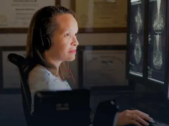
Teleradiology services leader since 2001. See how vRad AI is helping deliver faster, higher-quality care for 50,000+ critical patients each year.
Subspecialist care for the women in your community. 48-hour screenings. 1-hour diagnostics. Comprehensive compliance and inspection support.
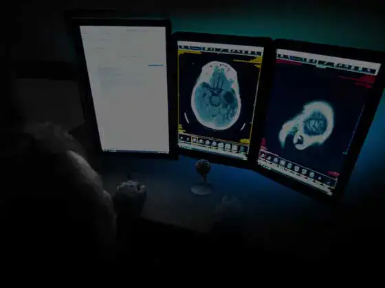
vRad’s stroke protocol auto-assigns stroke cases to the top of all available radiologists’ worklists, with requirements to be read next.

vRad’s unique teleradiology workflow for trauma studies delivers consistently fast turnaround times—even during periods of high volume.
vRad’s Operations Center is the central hub that ensures imaging studies and communications are handled efficiently and swiftly.
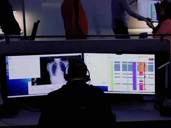
vRad is delivering faster radiology turnaround times for 40,000+ critical patients annually, using four unique strategies, including AI.
vRad is developing and using AI to improve radiology quality assurance and reduce medical malpractice risk.
Now you can power your practice with the same fully integrated technology and support ecosystem we use. The vRad Platform.
Since developing and launching our first model in 2015, vRad has been at the forefront of AI in radiology.
Since 2010, vRad Radiology Education has provided high-quality radiology CME. Open to all radiologists, these 15-minute online modules are a convenient way to stay up to date on practical radiology topics.
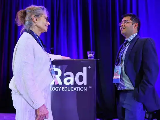
Join vRad’s annual spring CME conference, featuring top speakers and practical radiology topics.
vRad provides radiology residents and fellows with free radiology education resources for ABR boards, noon lectures, and CME.

Academically oriented radiologists love practicing at vRad too. Check out the research published by vRad radiologists and team members.

Learn how vRad revolutionized radiology and has been at the forefront of innovation since 2001.

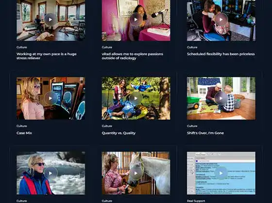
Explore vRad’s award-winning culture and what it’s like to work at vRad.
Meet vRad’s executive and medical leaders.
Visit the vRad blog for radiologist experiences at vRad, career resources, and more.

Get the latest news from vRad and top stories over the years.
Explore our practice’s reading platform, breast imaging program, AI, and more. Plus, hear from vRad radiologists about what it’s like to practice at vRad.

Ready to be part of something meaningful? Explore team member careers at vRad.

Journal Name: Clinical Imaging
Published: 11/01/2025
Journal Name: Clinical Imaging
Published: 02/01/2025
Journal Name: American College of Radiology
Published: 12/06/2023
Journal Name: Emergency Radiology
Published: 07/31/2023
Journal Name: Emergency Radiology
Published: 07/17/2023
Journal Name: Spine
Published: 01/01/2023
Journal Name: Journal of the American College of Radiology
Published: 11/01/2022
Journal Name: Journal of the American College of Radiology
Published: 05/01/2022
Journal Name: SPIE Medical Imaging
Published: 04/04/2022